
Journal of Multidisciplinary Dental Research
Volume: 4, Issue: 2, Pages: 46-51
Case Report
Fouziya Begum1, Uthappa M.A2, Rupesh P. L3, Basavaraj S Salagundi4, Bharat Kataraki5, Kongkona Bharadwaj6
1Postgraduate student, Coorg Institute of Dental Sciences, Virajpet
2Reader, Coorg Institute of Dental Sciences, Virajpet.
3Professor and Chair, Coorg Institute of Dental Sciences, Virajpet
4Professor and HOD, Coorg Institute of Dental Sciences, Virajpet
5Senior Lecturer, Coorg Institute of Dental Sciences, Virajpet
6Postgraduate student, Coorg Institute of Dental Sciences, Virajpet
Corresponding
Basavaraj
Professor and HOD, Coorg Institute of Dental Sciences, Virajpet
Ph: 9480905758
e-mail : [email protected]
Received Date:01 November 2018, Accepted Date:10 November 2018, Published Date:16 November 2018
This case report presents a patient who had been rehabilitated with implants supported mandibular fixed dental prosthesis and maxillary single unit and three units fixed partial dentures. Six implants were successfully placed in the mandibular arch and no complications occurred in the postoperative or maintenance periods. Meanwhile the maxillary arch was rehabilitated with fixed dental prosthesis taking esthetics and function into consideration. Four months after the mandibular prosthesis was delivered; which was found to be clinically, biologically, and mechanically stable, and the patient was satisfied with the esthetics and her ability to function. Direct metal laser sintered porcelain veneered restorations were used in the present report for its advantages of accurate restoration and marginal accuracy.
Full mouth rehabilitation always demands great attention and meticulous treatment planning. Patients often suffer from destructive dental caries and periodontal problems, which can lead to the early loss of teeth.1 Prosthetic rehabilitation of partial edentulism can be done with plenty of contemporary and conventional treatment modalities. Implant-supported prostheses have become widely preferred prosthetic approach and treatment choice because of their reliable functional and aesthetic results.2 The All-on-6 technique is specifically designed to use to use six implants, they do not typically require bone grafting and are an ideal solution for patients with areas of low bone density or volume in the posterior ridge region.3 Besides the implant treatment, meeting a patient's high esthetic demands depends on the acquirement of several biological and mechanical goals4. Porcelain veneered single crowns and fixed dental prostheses (FDPs) are well known for satisfying esthetics, biocompatibility, color stability, and resistance to wear.4 In recent years, direct metal laser sintered (DMLS) which is a digitalized metal casting technology, have been used as an alternative to conventional metal-ceramic fixed partial denture prostheses. It is relatively new; produces accurate restorations, simplified post processing procedures, free of porosity unlike conventional castings and improved electromechanical characteristics.5
Clinical report
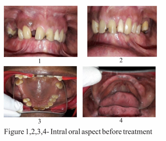
A 65 year old female patient reported to the department of prosthodontics with a chief complaint of poor esthetics and inability to chew because of missing teeth. After obtaining her medical, dental histories, she was examined clinically and radiographically. It was found that she had lost her all teeth in the lower jaw and many of the teeth in the upper jaw due to periodontal diseases and decay. Intraoral examination revealed absence of 12, 15, 16, 17, 26, 27 and the remaining anterior teeth present had poor esthetics.
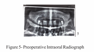
The radiographs were taken to evaluate the condition of the teeth that were present and the condition of the bone for implants. Patient was well informed and explained about the various treatment options like implant/tooth supported removable and implant supported fixed prostheses etc.
Considering the remaining teeth, periodontal tissues health and patient expectations/benefits, it was decided to perform implant supported mandibular fixed partial denture and individual crowns and fixed partial dentures for maxillary arch. The fixed prosthesis for mandibular arch was achieved in 3 sections- 3 unit FPD for the posterior segments and 6 units FPD as an anterior segment.
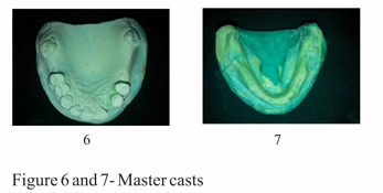
At the initial phase of first stage treatment protocol, border molding was done for the mandibular edentulous arch followed by maxillamandibular relation, which was mounted onto a semi adjustable articulator.
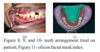
A temporary prosthesis was given to the patient for time being. Meanwhile, a silicone facial mask index was made for the mandibular arch to serve as a guide for the final restoration.
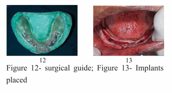
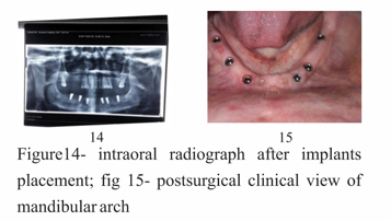
An open-flap surgery was performed and Adin dental implants were placed in the region of canine, first premolar and first molar of dimension 4.5×11.5mm.
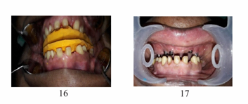
Meanwhile during the osseointegration period, the rehabilitation of the maxillary arch was carried out. A silicone putty index was prepared to serve as a guide for abutment occlusal reduction.
Figure 16- silicone index for abutment preparation; figure 17- view of prepared abutments
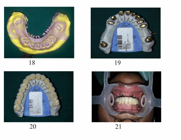
About 1.5 mm occlusal reduction was done. The preparation had a circumferential shoulder finish line design with a circumferential reduction of 1.5 mm and a total convergence of 6°. Impression was made using polyvinyl siloxane impression material and poured with Type IV die stone (Fuji rock, GC). DMLS posterior three-unit metal-ceramic complete coverage fixed partial dentures and individual crowns. Marginal integrity was checked and the final cementation was done using Type I glass ionomer cement.
Fig 11- impression of the prepared teeth of maxillary arch; Fig 19- cast with die spacer for fabrication of DMLS metal framework; Fig 20, 21- Rehabilitated maxillary arch
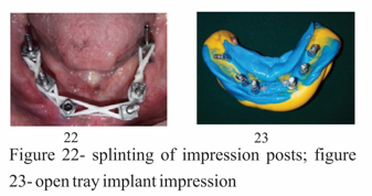
After 4 months of osseointegration of implants for mandible, second stage surgery was performed and healing abutments were placed. Two weeks later, impression coping were placed, splinted together intraorally with dental floss and an open tray impression was made using cold cure acrylic resin customized tray.
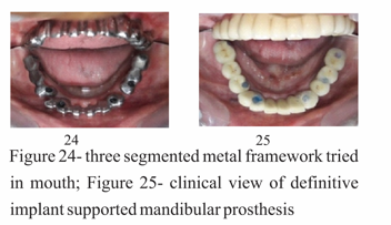
The metal frameworks obtained were tried and checked. Mandibular prosthesis with veneered porcelain was delivered.
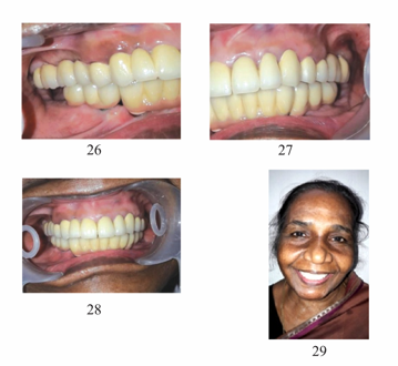
All the prosthetic screws were given an end most torque of 15N/cm4. The prosthetic screw access holes were sealed with putty and composite material. The occlusion was evaluated and adjusted to a mutually protected occlusion scheme keeping in mind the patient's centric relation
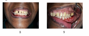
Figure 29- postoperative view
The patient was recalled for follow up visits every 6 weeks during the first 3 months, 6 months after the insertion of definitive prostheses. At 6 months, the prosthesis was removed and biological and mechanical complications were assessed. Further follow-ups visits were carried out at 12 and 18 months after delivery of the prosthesis.
DISCUSSION
Proper diagnosis and precise treatment planning are key to success for implant rehabilitation. Implant-supported FPD is a viable treatment option in the literature and has shown predictable long-term outcome. In the present patient, segmented fixed full-arch implant rehabilitation was selected for the definitive prosthetic design of the mandibular arch. The distribution of implants was followed according to the prosthodontic plan. The mandibular arch included a six-unit implant supported anterior FPD and two three-unit posterior implant supported FPDs on a total of six implants. Segmenting the FPDs into three FPD units was preferred over a one-piece prosthesis because of the advantages such as maintenance, decreased follow-up costs (in case of an abutment loss, porcelain fracture, or chipping), and improved distribution of occlusal and lateral loads6,7,8.Furthermore, segmenting it facilitates creation of embrasures, which enhances the natural appearance of the restoration.7 The implant supported mandibular prosthesis was screw retained considering the need for retrievability and easier management of complications.9 A finite element study suggested that placing 5-6 implants results in less stress distribution compared to those with 3 implants.10
Casting of alloys using the conventional technique is still widely practiced in dentistry Recent advancement in the use of computer-aided design and computer-aided manufacturing in dentistry, has led to tremendous change in casting of dental alloys5. Dr.Deckard and Beaman, introduced metal laser sintering which builds up the framework in a series of successively thin layers in the range of 0.02–0.06 mm11. DMLS results in highly accurate and well-detailed restorations can be fabricated using DMLS technique.12,13 Studies suggest that laser sintered metal crowns when compared with conventionally made cast crowns for the internal fit resulted in less marginal gap for laser sintered crowns of 65 µm when compared to the conventionally made crowns with a value of 150–125 µm14. So, based on the promising results obtained from various in vitro studies and in vivo studies for crowns and FPDs using DMLS technique, this study of giving crowns and fixed partial denture made from DMLS technique was undertake.
Regarding the definitive prosthetic plan for the maxilla in the present patient, several fixed options were considered. But because of the patient's desire to avoid removable prostheses and implants, a treatment sequence for the maxillary arch using a staged approach was carried out to rehabilitate the missing teeth and to improve the esthetics of the patient using PFM complete ceramic coverage restorations.
Conclusion
An excellent prognosis can be given for the present treatment, assuming continued favorable patient compliance with regard to oral hygiene maintenance. Implant supported fixed partial denture is a good alternative to the conventional complete dentures due to the better reconstruction of the patient's functional, phonation, and esthetic requirements. DMLS metal-ceramic fixed partial dentures have shown promising results during the observation period of 60 months by proving their clinical survival rate of 95.5% indicating greater clinical acceptability for use in day-to-day clinical practices.
Begum et al.,Full mouth rehabilitation with implant-supported fixed dental prosthesis – 'an all-on-6 clinical report'.2018:4(2);46-51
Subscribe now for latest articles, news.

